Before I started working in an embryology lab, I thought embryo development was pretty simple. Egg meets sperm. Embryo forms. Baby happens. Turns out there’s a lot more going on than that.
One of my favorite parts of learning embryology has been realizing just how much work these microscopic cells are doing in a very short amount of time. So if you’ve ever wondered what embryologists are actually looking at all day, here’s a basic breakdown of embryo development without the textbook language.
Quick note: Throughout this post, I’ll occasionally reference figures (images) to help visualize what’s happening. If I mention “Figure 1” or “Figure 2,” just scroll to the corresponding image and caption. Think of them as visual aids that go along with the explanation.
Fertilization
Everything just begins when an egg and sperm successfully come together. The day after fertilization, embryologists look for something called two pronuclei (2PN). These are essentially the genetic contributions from the egg and sperm before they merge (Figure 1).
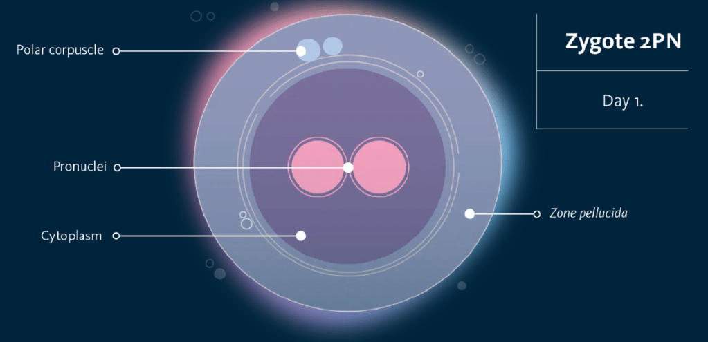
Seeing 2PN is one of the first signs that fertilization happened normally.
Cleavage Stage – The Copier
Over the next few days, the embryo starts dividing. One cell becomes two. Two become four. Four become eight (Figure 2).
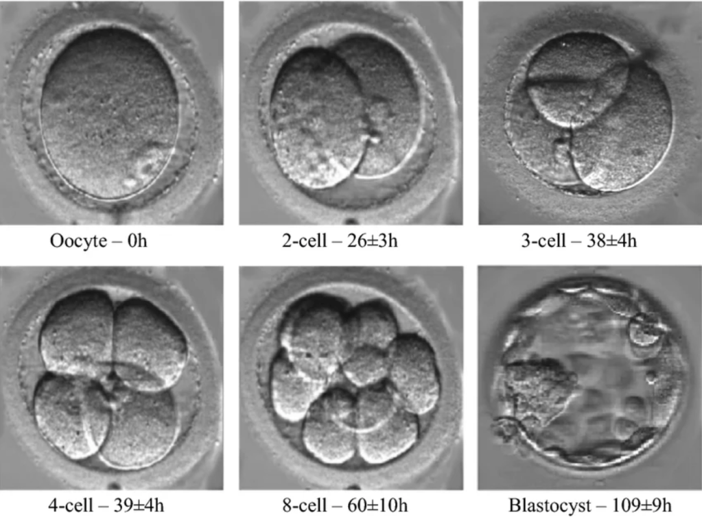
What’s fascinating is that the embryo isn’t really growing larger at this point. Instead, it’s dividing itself into smaller and smaller cells while staying roughly the same size. Embryologists watch how these cells divide and whether development is happening at an expected pace.
Compaction
Around Day 4, the embryo reaches a stage called compaction. This is when the individual cells start sticking tightly together and become harder to distinguish from one another. I always think of it as the point where all the cells decide to stop acting independently and start functioning as a team. They’re beginning to organize for what’s coming next.
Blastocyst Stage (Everybody Gets a Job)
By Day 5 or 6, some embryos reach the blastocyst stage. This is where things get really interesting. Instead of being a simple cluster of cells, the embryo starts separating into specialized groups. The inner cell mass is the group of cells that may eventually become the fetus. The trophectoderm is the layer that may eventually contribute to the placenta. Basically, these cells are already assigning responsibilities before they’re even visible without a microscope (Figure 2 – shows a blastocyst in the bottom right corner).
Embryo Grading (Thankfully Not a Report Card)
One thing I didn’t understand at first was embryo grading.
When embryologists assign grades, they’re describing what they see under the microscope. They’re looking at things like expansion, cell organization, and overall appearance.
Essentially, a grade helps communicate how an embryo looks at that moment. It isn’t a guarantee of success or failure. I like to think of grading as a snapshot rather than a prediction.
The Number = How Expanded the Blastocyst Is
The number tells us how far the blastocyst has expanded.
- 1 = Early Blastocyst (The embryo is just starting to form a fluid-filled cavity)
- 2 = Blastocyst (The cavity is larger and easier to identify)
- 3 = Full Blastocyst
The embryo has expanded further and is looking more organized. - 4 = Expanded Blastocyst
The blastocyst has grown larger and the zona pellucida (Figure 3) (the shell around the embryo) is thinning. - 5 = Hatching Blastocyst
The embryo has started breaking through its shell. - 6 = Hatched Blastocyst
The embryo has completely escaped the shell.
The First Letter = Inner Cell Mass
The first letter grades the Inner Cell Mass (ICM), which is the group of cells that may eventually become the fetus.
- A = Lots of tightly packed cells. Easy to identify.
- B = A good number of cells, but not packed quite as tightly.
- C = Fewer cells or less organized appearance (Figure 3).
The Second Letter (Trophectoderm)
The second letter grades the Trophectoderm (TE), which is the layer that may eventually become the placenta.
- A = Many cells forming a nice, organized layer.
- B = Moderate number of cells with decent organization.
- C = Fewer cells or a less cohesive appearance (Figure 3).
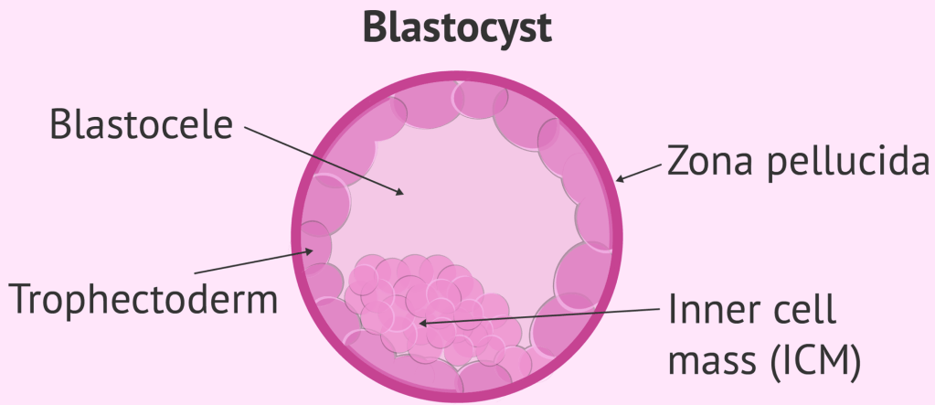
Embryologists use grading to compare embryos and help select which ones may have the best chance for transfer or freezing, but plenty of lower-graded embryos have resulted in healthy pregnancies, and plenty of beautiful-looking embryos haven’t implanted. (Biology loves reminding us that it doesn’t always follow the rules).
Why Embryology Is So Cool
What surprised me most about working in an embryology lab is how much development happens in just a few days.
In less than a week, a single fertilized cell can divide, organize itself, form specialized structures, and prepare for implantation. And all of that happens on a microscopic scale. Every day in the lab feels like a reminder that biology is somehow both incredibly complicated and incredibly efficient.
I Know…
Embryology can sound intimidating when it’s explained through textbooks and scientific papers (Trust me, I’ve spent enough time staring at diagrams wondering what I was looking at). At its core, embryology is really the story of how a few cells become something far more complex.
The more I learn in the lab, the more amazed I am by how much information is packed into those first few days of development. And honestly, that’s what keeps me coming back to learn more.
Living with Premature Ovarian Insufficiency (POI)
For the girls who feel too young for this, and too old to ignore it.



Leave a Reply